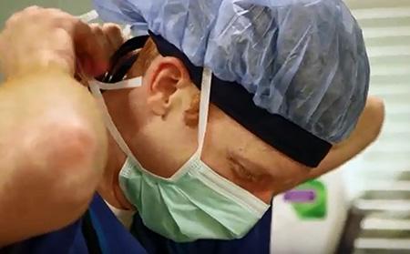
Novel technology from Duke researchers for identifying brain tumors during the course of surgery is showing promise.
The device, called “TumorID,” uses a nondestructive laser technology and machine learning to almost immediately classify a specimen as glioma, meningioma, pituitary adenoma, or nonneoplastic tissue while still in the operating room.
In the paper published on the cover of the Journal of Neurosurgery, TumorID was used to scan ex vivo specimens from 46 patients. The algorithm was trained to distinguish between the samples and classify them in near-real time.
“This work demonstrates the potential of TumorID to instantly identify brain tumor tissue in the operating room, an essential step toward a future where advanced sensors and ultraprecise, autonomously guided tools work together to maximize safe and optimal tumor resection to improve patient outcomes,” said Patrick Codd, MD, the paper’s senior author and leader of the Brain Tool Lab.
TumorID requires only half a second to collect data from the tissue without needing any dyes or tissue manipulation.
“The hope for the TumorID is to provide the surgical team with quantitative knowledge of point-by-point tissue diagnosis prior to tissue resection,” said Tanner Zachem, the paper’s first author. “This could allow the surgeons to tailor each brain and spine tumor resection by allowing for a more complete resection, while minimizing damage to adjacent normal tissues.”
Being able to quickly classify tissue types as being lesions or normal brain tissue during surgery is invaluable, especially for tumors like glioma that have notoriously invasive borders.
Zachem said the next step for TumorID is to investigate identifying key genetic mutations. In the future, Zachem said the hope is to also integrate TumorID into novel millimeter-sized robots to reach previously inaccessible locations.