Overview
Our research focuses on the development and clinical translation of novel quantitative magnetic resonance imaging (MRI) technology, to improve the understanding, diagnosis and treatment of diseases.
Clinical decisions currently rely heavily on invasive procedures, such as biopsies and surgical implants. There is a pressing clinical need to develop non-invasive, low-risk, time and cost efficient methods for improved diagnosis and treatment management. Our goal is to address this clinical need by developing an efficient MR imaging acquisition and post-processing framework to provide non-invasive, fast and comprehensive information for clinical decision support.
Our work focuses on:
-
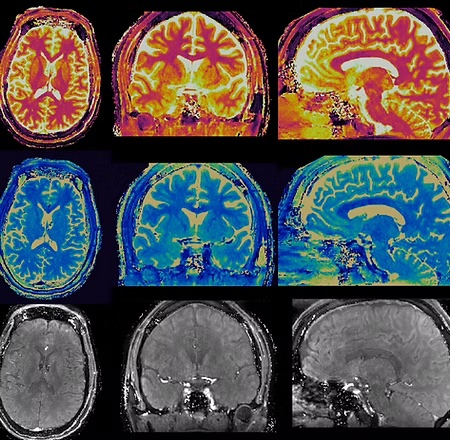
Magnetic resonance fingerprinting (MRF). MRF is a novel quantitative MR imaging technique developed in our lab which permits non-invasive quantification of multiple tissue properties within clinically feasible time.
-
Multi-parametric MR sequence design and optimization, image reconstruction, signal modeling and post-processing.
-
Clinical translation of MR fingerprinting in early detection and characterization of neurological diseases such as epilepsy, brain tumors, and multiple sclerosis.
Research
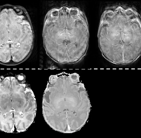
Comprehensive MR Fingerprinting for Infants and Young Children at Risk for Developmental Delay
Funding: NIH/NICHD R01
MPI: Dan Ma, Deanne E Wilson-Costello, Pew-Thian Yap
Collaborators: Duke University, University Hospitals Rainbow Babies & Children's, University of North Carolina at Chapel Hill
Development of Fast Diffusion Magnetic Resonance Fingerprinting of the Prostate to Avoid Unnecessary Biopsies
Optimal detection of clinically significant prostate cancer while avoiding overdiagnosis and unnecessary procedures
Funding: NIH/NCI R01
MPI: Leonardo Kaya Bittencourt, Dan Ma, Yong Chen
Collaborators: Duke University, University Hospitals Cleveland Medical Center
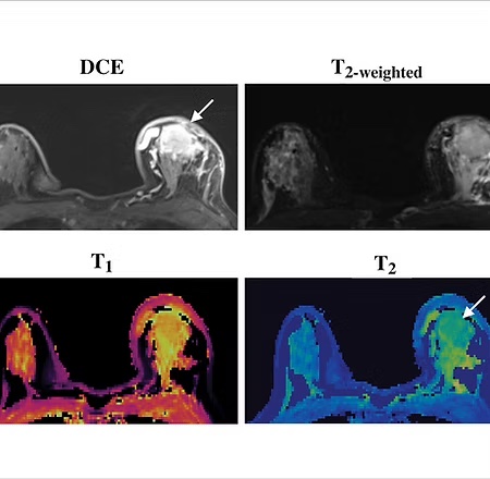
Development of Magnetic Resonance Fingerprinting (MRF) to Assess Response to Neoadjuvant Chemotherapy in Breast Cancer
Develop MRF methods to assess early response to neoadjuvant chemotherapy in women with breast cancer
Funding: NIH/NCI R01
MPI: Yong Chen, Dan Ma, Holly Marshall
Collaborators: Duke University, University Hospitals Cleveland Medical Center
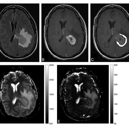
MR Fingerprinting Based Quantitative Imaging and Analysis Platform (MRF-QIA) for Brain Tumors
Academic industry partnership to translate quantitative imaging and analysis into the clinical workflow
Funding: NIH/NCI R01
MPI: Dan Ma, Chaitra Badve, Christos Davatzikos
Collaborators: Siemens Healthineers, Duke University, University Hospitals Cleveland Medical Center, University of Pennsylvania
Making the Invisible Visible
A multi-scale approach integrating MR imaging, tissue modeling, tissue imaging and deep learning to detect and characterize cortical pathology
Funding: UK Research and Innovation/ MRC
MPI: Derek Jones (contact), Dan Ma, Mark Griswold, Daniel Alexandar
Collaborators: Duke University, Cardiff University, University College London, University of Leeds
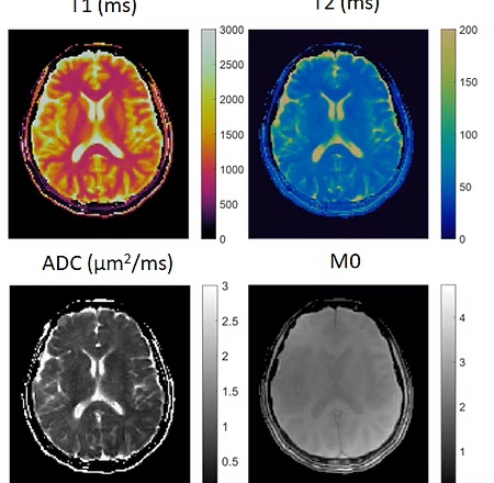
Clinically Feasible MR Fingerprinting Imaging Framework
- Simultaneous T1 and T2 mapping with whole brain coverage and isotropic image resolution
- Sequence optimization using quantum optimization algorithms
- Low rank subspace image reconstruction
- Partial volume analysis and tissue segmentation
Funding: Siemens Healthineers, NIH/NIBIB R21
Collaborators: Siemens Healthineers, Microsoft Quantum Team
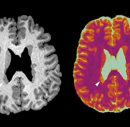
MR Fingerprinting in Epilepsy
Epilepsy affects 65 million people worldwide; approximately 30% of them do not respond to medications but can be cured by surgery. Focal cortical dysplasia, a major pathology for medically intractable epilepsies, are frequently missed by visual analysis of the conventional MRI, making surgical treatment very difficult. Here we propose to develop and validate novel, noninvasive and quantitative MRI acquisition and post-processing techniques, in order to guide epilepsy surgery and make more patients seizure-free.
Funding: NIH/NINDS R01
MPI: Dan Ma, Irene Wang
Collaborators: Duke University, Cleveland Clinic Epilepsy Center
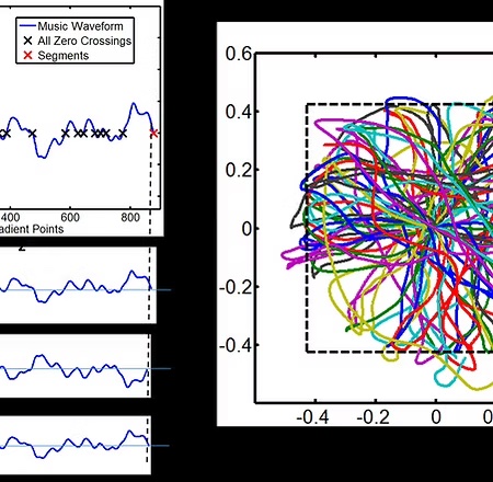
A Framework to Design 3D MRF Scans and Reduce Patient Anxiety (MRF Music!)
Loud noise during MRI scans is the leading cause of patients’ anxiety, but the origin of this loud noise, mainly fast-switching fields, is also an essential component to generate images. Previous methods rely almost solely on slowing down the switching field to reduce the noise, resulting in reduced scan efficiency. We propose a general framework that could resolve this longstanding conflict by changing the sound of the MRI scan to music while simultaneously providing multiple quantitative tissue properties with high scan efficiency.
Funding: NIH/NIBIB R21 Trailblazer
PI: Dan Ma
Collaborators: Duke University
Personnel
Dan Ma, PhD
Principal Investigator
Associate Professor in Neurosurgery
Associate Professor in Biomedical Engineering
Duke University
dan.ma@duke.edu
ORCID: 0000-0003-1664-9579
Publications: Google Scholar

Shahrzad Moinian, PhD
Senior Research Associate
shahrzad.moinian@duke.edu
MS in Computer Science, University of Queensland, Australia
PhD in Computational Neuroimaging, University of Queensland, Australia
Shahrzad Moinian specializes in medical image acquisition and analysis with a particular interest in developing artificial intelligence-based methods for the reconstruction and analysis of magnetic resonance imaging across various clinical applications. Her doctoral research led to the development of the MR fingerprinting residual analysis framework, a novel method for in vivo detection and characterization of microstructural variations in the human cerebral cortex. Currently, Shahrzad's work focuses on advancing multi-dimensional MR fingerprinting acquisition and analysis for clinical translation into body imaging, aiming to enhance diagnostic capabilities and patient care.
Outside of work, she enjoys woodworking projects, playing piano, and participating in social soccer.
Kewei Yan, Ph.D.
Postdoc Associate
kewei.yan@duke.edu
M.S. in Biostatistics, University of Connecticut, CT
Ph.D. in Computer Science University of North Carolina at Charlotte, NC
Kewei Yan's research interests covering High-performance Computing (HPC) and Machine Learning (ML). The dissertation is an approach of surrogate-assisted in-situ analysis for efficient data analysis from scientific simulation.
In his free time, Kewei enjoys maintaining aquarium, painting, playing musical instrument, and playing video games.

Rhea Adams
PhD student in Biomedical Engineering
richard.j.adams@duke.edu
BS in Biomedical Engineering, University of Houston

Eunate Alzaga
PhD student in Biomedical Engineering at Duke
eunate.alzaga@duke.edu
BS in Biomedical Engineering, University of Rochester
Eunate's research focuses on the development of MRF-based deep learning algorithms. In her free time, she enjoys watching movies, reading novels, traveling, and trying new restaurants.
Rory (Tianhe) Wu
PhD student in Biomedical Engineering at Duke
rory.wu@duke.edu
BS in Applied Mathematics & BA in Biology., Emory University
In his free time, Rory enjoys playing tennis and watching movies.
Yanfei Xiong
PhD student in Biomedical Engineering at Duke
yanfei.xiong@duke.edu
MS in Physics, University of Science and Technology of China
BS in Applied Physics, Shandong University
Yanfei enjoys watching tennis matches, baking, traveling, and always has a passion for exploring new experiences and adventures.

Zhicheng (Russell) Wu
MS student in Biomedical Engineering at Duke
Outside of Duke, Zhicheng enjoys soccer, Counter-Strike, and film.
Alumni
Jessie Sun
BSE in Materials Science & Engineering, University of Pennsylvania
Zhilang Qiu
Research Scientist at McLean Hospital, Harvard School of Medicine;
PhD in Pattern Recognition and Intelligent System, University of Chinese Academy of Sciences, Beijing, China
Siyuan (Cindy) Hu
Cardiac MRI Scientist at GE HealthCare;
PhD student in Biomedical Engineering;
BS in Biomedical Engineering, Case Western Reserve University;
2024 Doctoral Excellence
Zheyuan (Jason) Hu
Graduate student researcher at UCLA;
BS student in Applied Mathematics and Biomedical Engineering
Christina MacAskill
MD-PhD student in Biomedical Engineering;
BS in Biomedical Engineering, Case Western Reserve University
Walter Zhao
MD-PhD Student
PhD in Biomedical Engineering at Case Western Reserve University
MD student at Case Western Reserve University
News


Contact
The Quantitative MR Imaging Lab is seeking to hire graduate students and postdoctoral fellows. Interested candidates should send their CV to Dan Ma, PhD, at dan.ma@duke.edu.
Location
445 Davison Building
Duke University
Durham, NC 27708